A chest X-ray can reveal signs of a pulmonary embolism, and understanding key facts is important.
Diagnosing Pulmonary Embolism: A Look at Imaging Techniques
Pulmonary embolism (PE), a potentially life-threatening condition, can be challenging to diagnose. However, advancements in medical imaging have made it easier to identify this condition and manage it effectively. Here's a rundown of the key imaging tests used in diagnosing PE.
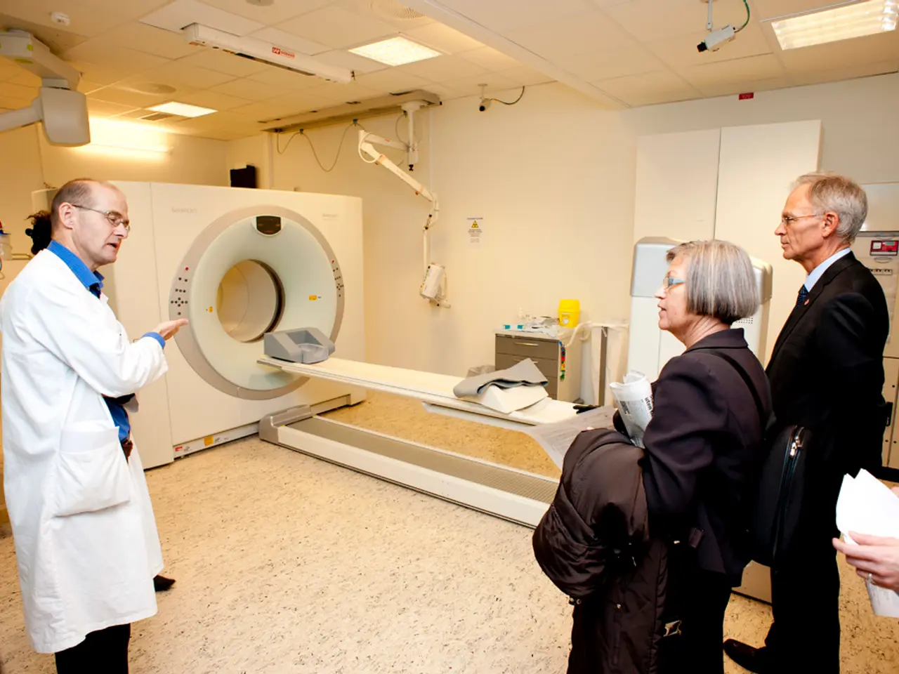
CT Pulmonary Angiography (CTPA) is currently the preferred and most definitive imaging test for PE. This test directly visualizes intraluminal filling defects (blood clots) in the pulmonary arteries, especially at the segmental and more proximal levels. Multidetector CT improves detection of smaller, subsegmental emboli. Its sensitivity and specificity are approximately 85-90% and 90-95%, respectively, making it a reliable tool for diagnosing PE [2][3][4].
Pulmonary Angiography involves injecting contrast dye into the pulmonary arteries via a catheter to detect clots directly. While it is a definitive diagnostic test, it is more invasive and less commonly used today due to advances in CT imaging [1].
Ventilation-Perfusion (V/Q) Scan assesses the distribution of air flow (ventilation) and blood flow (perfusion) in the lungs. A mismatch — normal ventilation but decreased perfusion — suggests PE. It is a non-invasive alternative, especially for patients who cannot receive contrast dye [1][4].
Ultrasound, specifically venous ultrasound of the lower extremities, is commonly used to detect deep vein thrombosis (DVT), a frequent source of PE. While it does not image pulmonary arteries, it helps support the diagnosis of PE by finding clots in leg veins and can guide management indirectly [1][4].
Echocardiography (ultrasound of the heart) can identify right ventricular strain or dysfunction caused by obstructed pulmonary blood flow, aiding presumptive diagnosis especially in unstable patients when CT may be unsafe [1][4].
In addition, advanced techniques like dual-energy CT with AI-assisted quantification of lung perfusion defects can better evaluate embolic burden and correlate with clinical severity, potentially enhancing diagnosis and management [5].
In summary, CT pulmonary angiography is the frontline imaging modality for diagnosing PE by directly showing pulmonary artery clots, while ultrasound helps detect contributing peripheral clots. The ventilation-perfusion scan or angiography serves as alternatives or confirmatory tests when CT is contraindicated or inconclusive. Echocardiography provides functional assessment in critical cases. Each imaging modality contributes unique diagnostic information to confirm or rule out pulmonary embolism.
System-based diagnostics have revolutionized the medical world, and this is particularly true for cardiovascular and respiratory conditions such as pulmonary embolism (PE). Advanced imaging technologies have made PE diagnosis more accessible and effective. CT Pulmonary Angiography (CTPA), with its high sensitivity and specificity, is commonly used due to its ability to visualize intraluminal filling defects. Pulmonary Angiography, though definitive, is less often utilized today due to advancements in CT imaging. A Ventilation-Perfusion (V/Q) Scan, a non-invasive alternative, assesses lung airflow and blood flow, while ultrasound aids in detecting deep vein thrombosis (DVT), a frequent PE source. Echocardiography, on the other hand, offers functional assessment in critical cases by identifying right ventricular strain. Hence, each imaging modality plays a unique role in PE diagnosis, contributing valuable information to either confirm or rule out this potentially life-threatening medical-condition.


