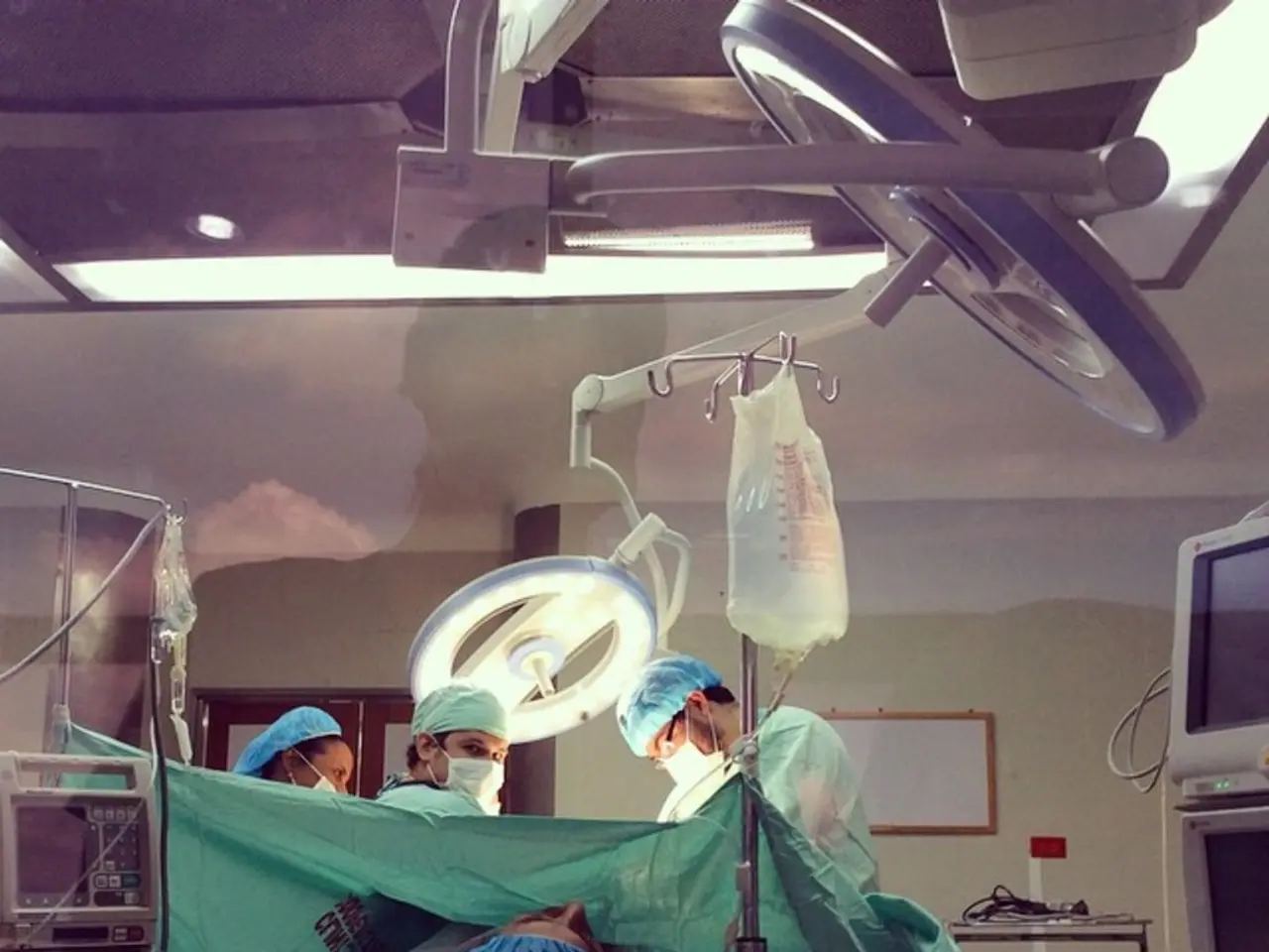
Innovative Camera Technology Now Capable of Penetrating Human Bodies
A groundbreaking camera technology is making waves in the medical field, promising to revolutionise the way physicians diagnose and treat diseases. This technology, currently exemplified by advanced imaging systems such as the MARS spectral X-ray scanner from the University of Canterbury and MIT's multiphoton photoacoustic microscope, offers unprecedented 3D visualisation of soft tissues and brain structures with high resolution.
Applications
The MARS spectral X-ray scanner creates 3D colour images of the human body using spectral imaging combined with Medipix3 particle detector technology from CERN. This technology can distinguish tissues like bones, fat, water, and cartilage, aiding in non-invasive diagnosis of cancers and blood diseases, potentially replacing invasive surgery.
MIT's multiphoton photoacoustic microscope, on the other hand, images brain tissue up to five times deeper than previous methods without dyes or genetic markers. It uses three-photon excitation and sound wave detection to reveal deep brain structures and metabolites critical to neuroscience research, with future prospects for surgical guidance and brain disease studies.
Scientific Principles
The MARS scanner uses spectral X-ray imaging and particle counting, enabling high-resolution soft tissue contrast by capturing the energy spectrum at each pixel. The MIT system, in contrast, combines ultrashort infrared light pulses to generate microscopic thermal expansion in tissue, producing sound waves detected by ultrasound microphones. This three-photon photoacoustic imaging method reduces light scattering, allowing deeper, high-resolution imaging without invasive labels or dyes.
Future Prospects
These technologies promise routine clinical use for early disease detection, treatment monitoring, and brain research enhancement. Ongoing improvements aim to increase imaging depth, resolution, and affordability, for example, with advances in light sheet microscopy like MBF Bioscience’s SLICE system that provides accessible, affordable 3D imaging for biological samples, potentially complementing these deeper tissue imaging techniques.
Large-scale imaging datasets, such as from UK Biobank, leverage such technological advances to transform global health research by rapidly analyzing organ size, shape, and composition.
A New Paradigm in Medical Visualization
In summary, these revolutionary camera systems combine physics, engineering, and computational advances to see through human tissue in unprecedented detail and depth. They open new frontiers in medical diagnostics, neuroscience, and biological research by providing non-invasive, label-free, three-dimensional views inside the human body and brain.
The technology represents a new paradigm in medical visualization, where the boundaries between external observation and internal examination blur. Future iterations aim to address limitations such as maximum penetration depth, resolution decrease with tissue depth, and limited ability to penetrate certain dense tissue types.
In the clinical settings of tomorrow, seeing through the human body may become as routine as taking a pulse is today. Integration with existing medical imaging systems is necessary for clinical use. The new technology provides clear location information for the light source, whereas conventional cameras do not.
This technology represents a philosophical advancement, pushing the boundaries of what is considered observable and known in medicine. Clinical trials are required to validate the safety and efficacy of the technology, and regulatory approval is necessary for its reach in clinical use. The technology can detect an optical endomicroscope through sheep lung tissue, and the research team is exploring the use of near-infrared wavelengths to penetrate tissue more effectively.
The ultimate promise of this technology isn't just better images—it's fundamentally better medicine, where observation becomes less disruptive to the observed. The technology offers the possibility that physicians can navigate patients' bodies with greater precision and confidence, potentially leading to early disease detection, precise treatment delivery, and reduced procedural invasiveness. In the future, physicians may routinely "see" through tissue as easily as they now listen through a stethoscope.
Read also:
- Americans Lose Insurance Under New Tax Legislation, Affecting 10 Million Citizens
- TMJ Osteoarthritis: A Comprehensive Look at Its Nature
- Symptoms, Diagnosis, and Management Strategies for High-functioning ADHD
- Struggles of Advanced Breast Cancer Patients in Receiving Necessary Treatments Due to Conflicts and Bias


