Monitoring Lung Performance through innovative Scanning Technique
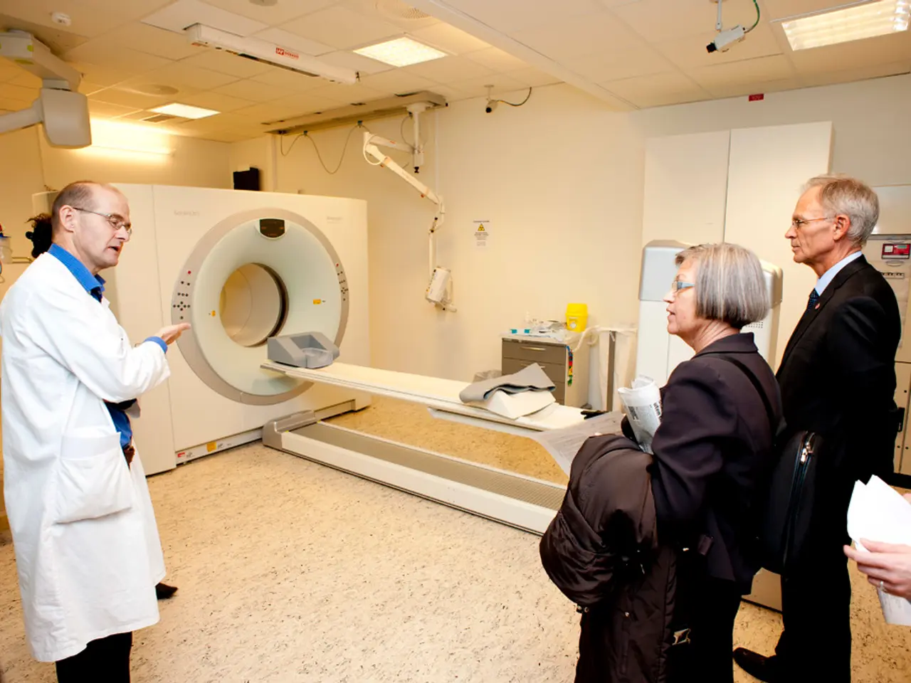
A groundbreaking new lung scanning method developed by researchers at Newcastle University is set to revolutionize the way respiratory diseases are diagnosed and managed. This innovative technique, which uses a special gas during MRI scans to visualize airflow through the lungs, offers real-time insights into lung function and treatment effects.
The new method has been shown to be highly effective in monitoring respiratory conditions like asthma, COPD, and lung transplants. By providing detailed, non-invasive assessments of lung structure and function over time, the technique improves clinical monitoring and enables personalized treatment adjustments based on precise lung changes.
The scanning technique works by quantifying how much of the lung is functioning well versus poorly, providing valuable information for assessing the severity of respiratory diseases. When a patient uses an asthma inhaler during the scan, doctors can see which parts of the lungs are better able to move air after the medication takes effect.
One of the most promising aspects of the new method is its ability to detect changes in lung function earlier than current methods allow, particularly in lung transplant recipients. The scans can reveal early signs of chronic rejection in these patients, potentially enabling doctors to intervene sooner and improve outcomes.
The research, funded by the Medical Research Council and The Rosetrees Trust, represents a significant step forward in respiratory medicine. The technique offers a clearer understanding of how air moves through the lungs, which could enhance care for patients with various respiratory challenges.
Professor Andrew Fisher, a co-author of the study, expressed hope that the technology will eventually be incorporated into routine care for lung transplant patients. The scans could help initiate treatment sooner for a variety of respiratory diseases, potentially preventing further complications.
The new lung scanning method also has potential for broader use in managing various lung diseases beyond lung transplants and common respiratory conditions like asthma and COPD. The scans reveal uneven ventilation in the lungs, helping to pinpoint problematic areas.
In clinical trials, the scans have proven valuable by measuring improvements seen after patients used a common bronchodilator, salbutamol. The technique offers a safer alternative to CT scans, which involve radiation, and provides more detailed information than standard pulmonary function tests.
The new MRI lung scan technique provides a safe, detailed, and repeatable imaging modality that enhances monitoring of chronic respiratory diseases and aids in optimizing treatment decisions. Further peer-reviewed clinical data may provide more precise effectiveness measures as this technology advances. This innovative approach offers new hope for patients with a variety of respiratory challenges, potentially transforming the way lung diseases are diagnosed and managed.
- The new lung scanning method, which uses special gas during MRI scans, can visualize airflow through the lungs, revolutionizing diagnosis and management of medical-conditions like chronic-kidney-disease, type-2-diabetes, and neurological-disorders that are often associated with poor cardiovascular-health and skin-conditions.
- This technique, highly effective in monitoring respiratory-conditions, offers real-time insights into lung function and treatment effects, allowing non-invasive assessments of lung structure and function over time.
- Besides tracking respiratory-conditions like asthma, COPD, and lung transplants, the method has potential for managing chronic-diseases like rheumatoid-arthritis, which can affect eye-health and fitness-and-exercise routines.
- The scans can reveal early signs of chronic rejection in lung transplant recipients, potentially enabling physicians to intervene sooner and improve outcomes, enhancing health-and-wellness overall.
- Technology in this MRI lung scan technique offers a safer alternative to CT scans, which involve radiation, and provides more detailed information than standard pulmonary function tests, thus being a crucial aid in nutrition-related evaluation.
- Scientists believe that further research on this innovative approach could lead to its incorporation into routine care for managing various lung diseases, impacting fitness-and-exercise, cardiovascular-health, and even eye-health.
- The new method gives a distinct advantage in catching anomalies like skin-conditions early, transforming the way lung diseases are diagnosed and managed, ultimately fostering health-and-wellness on a broader scale.
- Considering the clear benefits when implemented and the potential impact on patient care, investment in technology and scientific research for health-and-wellness initiatives, including fitness-and-exercise, nutrition, and eye-health, is more important than ever.


